

Focuses Light On The Retina
To understand Keratoconus, nosotros must first understand how the eye enables us to see, and what part the cornea plays in this process.
View Video
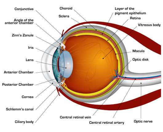
Low-cal rays enter the middle through the cornea, the clear front "window" of the eye. The cornea'due south refractive ability bends the light rays in such a way that they pass freely through the pupil the opening in the center of the iris through which lite enters the eye.
The iris works like a shutter in a photographic camera. It has the ability to enlarge and shrink, depending on how much light is inbound the center.
After passing through the iris, the light rays laissez passer thru the heart's natural crystalline lens. This clear, flexible structure works like the lens in a photographic camera, shortening and lengthening its width in order to focus low-cal rays properly.
Light rays laissez passer through a dense, transparent gel-like substance, called the vitreous that fills the globe of the eyeball and helps the eye hold its spherical shape.
In a normal eye, the light rays come to a sharp focusing bespeak on the retina. The retina functions much like the film in a camera. It is responsible for capturing all of the light rays, processing them into light impulses through millions of tiny nerve endings, and so sending these calorie-free impulses through over a meg nervus fibers to the optic nervus.
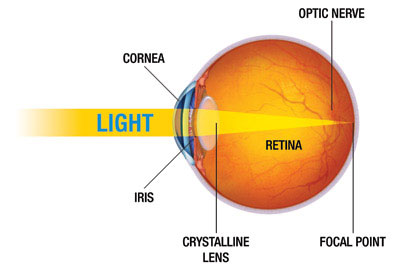
Considering the keratoconus cornea is irregular and cone shaped, light rays enter the centre at different angles, and do non focus on 1 point the retina, simply on many different points causing a blurred, distorted image.
In summary, the cornea is the articulate, transparent front covering which admits light and begins the refractive process. It also keeps foreign particles from entering the heart.
The pupil is an adjustable opening that controls the intensity of low-cal permitted to strike the lens. The lens focuses light through the vitreous humor, a clear gel-like substance that fills the dorsum of the eye and supports the retina.
The retina receives the paradigm that the cornea focuses through the centre'due south internal lens and transforms this paradigm into electrical impulses that are carried by the optic nerve to the brain. Nosotros can tolerate very large scars on our bodies with no business except for our vanity. This is not so in the cornea. Even a minor scar or irregularity in the shape can impair vision. No matter how well the balance of the eye is functioning, if the cornea is scarred, clouded or distorted, vision will be affected.
In keratoconus, the irregular shape of the cornea does not allow it to practise its job correctly, leading to baloney of the image it passed to the retina and transmitted to the brain.
 THE CORNEA
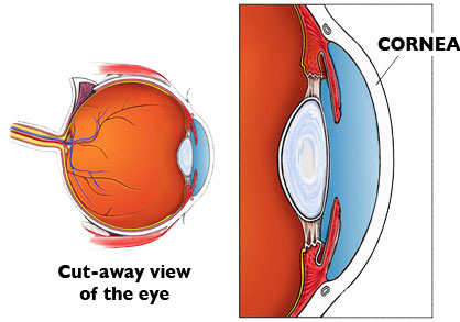
The middle is enclosed by a tough white sac, the sclera. The cornea is the transparent window in this white sac which allows the objects you lot are looking at to be carried in the form of low-cal waves into the interior of the eye.
The surface of the cornea is where light begins its journeying into the center. The cornea's mission is to get together and focus visual images. Considering it is out front, like the windshield of an auto, it is discipline to considerable corruption from the exterior world.
The cornea is masterfully engineered so that merely the near expensive manmade lenses can friction match its precision. The smoothness and shape of the cornea, as well as its transparency, is vitally important to the proper functioning of the eye. If either the surface smoothness or the clarity of the cornea suffers, vision volition be disrupted.
CORNEAL LAYERS
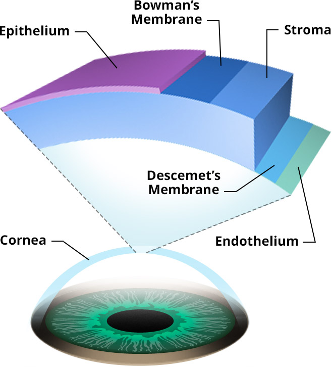 Although actualization to exist i clear membrane, the cornea is composed of five distinct layers of tissue, each with its own role.
- Epithelium is the thin outermost layer of fast-growing and hands-regenerated cells.
- Bowman'southward layerconsists of irregularly-arranged collagen fibers and protects the corneal stroma. Information technology is viii to xiv microns thick.
- Stroma, the transparent center and thickest layer of the cornea is made up of regularly-bundled collagen fibers and keratocytes (specialized cells that secrete the collagen and proteoglycans needed to maintain the clarity and curvature of the cornea)
- Descemet's membrane is a thin layer that serves as the modified basement membrane of the corneal endothelium.
- Endothelium is a single layer of cells responsible for maintaining proper fluid balance between the aqueous and corneal stromal compartments keeping the cornea transparent.

Focuses Light On The Retina,
Source: https://nkcf.org/about-keratoconus/how-the-human-eye-works/
Posted by: webbageres.blogspot.com
0 Response to "Focuses Light On The Retina"
Post a Comment